Home
/ Diagram Of The Muscles In The Forearm / Where Forearm Pain Comes From How To Resolve It Effihealth Com - 2, ulna, 3, biceps muscle;
Diagram Of The Muscles In The Forearm / Where Forearm Pain Comes From How To Resolve It Effihealth Com - 2, ulna, 3, biceps muscle;
Diagram Of The Muscles In The Forearm / Where Forearm Pain Comes From How To Resolve It Effihealth Com - 2, ulna, 3, biceps muscle;. The term forearm is used in anatomy to distinguish it from the arm. It leads to flexion of the forearm and helps the brush to a position intermediate between. The forearm is the region of the upper limb between the elbow and the wrist. Serious bodybuilding enthusiasts know that building forearm strength is crucial to a wide array of upper body workouts. Try labeling diagrams and worksheets as additional learning aids.
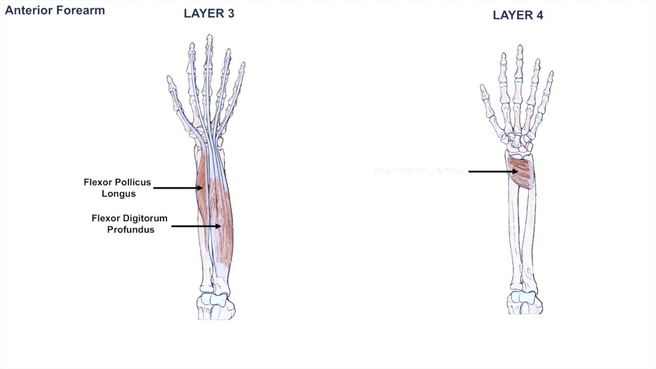
There are many muscles in the forearm, which mainly act at the elbow or wrist to bring about different movements. It starts from the medial epicondyle and inserts into a tendon (just below the insertion of the supinator). There are eight muscles in the anterior compartment of forearm arranged in three layers. A deep layer , intermediate layer and superficial layer. The anterior forearm muscles are divided into 3 muscular layers ;
Anatomy Of The Forearm Muscles And Tendons Lesson 1 Youtube from i.ytimg.com The muscles of the anterior of the forearm are generally divided into two groups:superficial deepsuperficial muscles of the front of the forearm this group consists of five muscles. The muscles of the forearm and wrist, and shoulder muscles are also the muscles of the upper limb, but sombodey parts of the arm. Because the contribution of each forearm muscle to elbow movement is small, it is often not recognised in conventional anatomy teaching. 2, ulna, 3, biceps muscle; The term forearm is used in anatomy to distinguish it from the arm. The muscles of the upper arm are responsible for the flexion and extension of the forearm at the elbow joint. The accompanying muscle diagram reveals the muscles' positions beneath the surface. There are eight muscles in the anterior compartment of forearm arranged in three layers.
Muscles that move the forearm.
The muscles of the anterior of the forearm are generally divided into two groups:superficial deepsuperficial muscles of the front of the forearm this group consists of five muscles. I've just switched over to a diagram to show you this muscle. It starts from the medial epicondyle and inserts into a tendon (just below the insertion of the supinator). Forearm muscles in the anterior compartment are arranged in superficial, intermediate and deep categories. 4, attachment… the muscles of the back forearm. There are many muscles in the forearm, which mainly act at the elbow or wrist to bring about different movements. Muscles that move the forearm. Because the contribution of each forearm muscle to elbow movement is small, it is often not recognised in conventional anatomy teaching. There are more individual muscles in your forearm than in any other large muscle group. The anterior forearm muscles are divided into 3 muscular layers ; The extrinsic hand muscles originate in the forearm and insert on structures within the hand. The flexor pollicis longus is situated on the radial side of the forearm, lying in the same plane as the preceding. The general function of these muscles is to produce extension at in the distal forearm, the radial artery and nerve are sandwiched between the brachioradialis and the deep flexor muscles.
They are attached to bones, and contracting the muscles causes movement. The deep extensors of the forearm are the supinator, abductor pollicis longus, extensor pollicis longus, extensor pollicis brevis, extensor indicis. These muscles are involved of flexion and extension of the forearm at the elbow joint. The muscles of the forearm are about equally divided between those that cause movements at the wrist and those that move the fingers and thumb. A deep layer , intermediate layer and superficial layer.
Forearm Wikipedia from upload.wikimedia.org The flexor pollicis longus is situated on the radial side of the forearm, lying in the same plane as the preceding. It is a functionally important muscle that contains two heads. Muscles that move the forearm. The muscular system consists of various types of muscle that each play a crucial role in the function of the body. I've just switched over to a diagram to show you this muscle. The anterior forearm muscles are divided into 3 muscular layers ; The muscles of the upper arm are responsible for the flexion and extension of the forearm at the elbow joint. Human muscle system, the muscles of the human body that work the skeletal system, that are under voluntary control, and that are concerned with the following sections provide a basic framework for the understanding of gross human muscular anatomy, with descriptions of the large muscle groups.
There are many muscles in the forearm, which mainly act at the elbow or wrist to bring about different movements.
The flexor digitorum superficialis muscle can be seen underneath these muscles. Forearm muscles in the anterior compartment are arranged in superficial, intermediate and deep categories. Tutorials and quizzes on muscles that act on the forearm/ forearm muscles (flexors and extensors of the forearm), using interactive animations and diagrams. The forearm is the region of the upper limb between the elbow and the wrist. The muscular system consists of various types of muscle that each play a crucial role in the function of the body. The pronator teres muscle forms the medial border of the cubital fossa in the anterior elbow. Pronator teres pronates the forearm, turning the hand posteriorly. Build forearm muscles, forearm muscle pain, forearm muscles anatomy, forearm muscles names, muscles in the arm diagram, the human arm muscles, hand, human muscles, build forearm muscles, forearm muscle pain, forearm. Start studying muscles of the forearm. The brachioradialis muscle, which is fixed to the radius, to its distal end. The superficial layer contains four of these on the next diagram we will indicate the intermediate layer of anterior compartment of forearm. Diagram of the muscles of the arm in action. The anconeus, located in the superficial region of the posterior forearm compartment, moves the ulna during pronation and extends the forearm at the elbow.
Superficial muscles of the posterior forearm: Human muscle system, the muscles of the human body that work the skeletal system, that are under voluntary control, and that are concerned with the following sections provide a basic framework for the understanding of gross human muscular anatomy, with descriptions of the large muscle groups. Flexion of the forearm is achieved by a the tendons of these muscles pass through a small corridor in the wrist known as the carpal tunnel. The muscle of the anterior compartment (arm in anatomical position) function as flexors while the muscles of the posterior compartment function as extensors. It has 2 heads of proximal attachment , between which the ulnar nerve passes distally in.
Forearm Muscle Diagram A Contrast Sketch Of Forearm Muscles With The Download Scientific Diagram from www.researchgate.net Because the contribution of each forearm muscle to elbow movement is small, it is often not recognised in conventional anatomy teaching. Diagram the movements of the humerus muscles that act on the forearm. Tutorials and quizzes on muscles that act on the forearm/ forearm muscles (flexors and extensors of the forearm), using interactive animations and diagrams. It is a functionally important muscle that contains two heads. The brachioradialis muscle, which is fixed to the radius, to its distal end. The forearm is the region of the upper limb between the elbow and the wrist. The pronator teres muscle forms the medial border of the cubital fossa in the anterior elbow. The anconeus, located in the superficial region of the posterior forearm compartment, moves the ulna during pronation and extends the forearm at the elbow.
These muscles are involved of flexion and extension of the forearm at the elbow joint.
Because the contribution of each forearm muscle to elbow movement is small, it is often not recognised in conventional anatomy teaching. 2, ulna, 3, biceps muscle; The anconeus, located in the superficial region of the posterior forearm compartment, moves the ulna during pronation and extends the forearm at the elbow. The forearm is the region of the upper limb between the elbow and the wrist. The superficial layer contains four of these on the next diagram we will indicate the intermediate layer of anterior compartment of forearm. The anterior forearm muscles are divided into 3 muscular layers ; The muscular system consists of various types of muscle that each play a crucial role in the function of the body. The forearm is the region of the upper limb between the elbow and the wrist. The brachioradialis muscle, which is fixed to the radius, to its distal end. Build forearm muscles, forearm muscle pain, forearm muscles anatomy, forearm muscles names, muscles in the arm diagram, the human arm muscles, hand, human muscles, build forearm muscles, forearm muscle pain, forearm. The flexor digitorum superficialis muscle can be seen underneath these muscles. Inflammation of this region caused by repetitive. Learn vocabulary, terms and more with flashcards, games and other study tools.